|
Introduction
Bloodstream
infections (BSIs) are a significant contributor to
morbidity and mortality among critically ill
patients and continue to impose a considerable
burden on healthcare systems. The risk is
particularly heightened in intensive care units
(ICUs), where factors such as compromised host
defenses, frequent use of invasive medical
devices, prolonged hospital stays, and extensive
exposure to broad-spectrum antimicrobials
predispose patients to systemic infections [1,2].
Collectively, these conditions create a highly
vulnerable environment that facilitates microbial
invasion and dissemination into the bloodstream.
The microbial
epidemiology of BSIs in ICU settings is highly
variable and often reflects local patient
characteristics, antimicrobial usage patterns, and
infection control practices. In recent years,
Gram-negative bacilli have emerged as the
predominant pathogens in many critical care
settings, especially species such as Klebsiella
pneumoniae, Acinetobacter baumannii,
and Pseudomonas aeruginosa. These
organisms are frequently associated with multidrug
resistance, significantly complicating therapeutic
management. At the same time, Gram-positive
organisms, including methicillin-resistant Staphylococcus
aureus (MRSA) and Enterococcus
spp., continue to contribute substantially to the
burden of bloodstream infections. Additionally,
fungal pathogens—particularly Candida
species—have gained increasing importance in
patients with prolonged ICU stays and multiple
comorbidities [3–5].
A key challenge in
the management of BSIs is the rapid and ongoing
rise in antimicrobial resistance (AMR). The
empirical use of broad-spectrum antibiotics,
although essential for early sepsis management,
exerts considerable selective pressure that
facilitates the emergence and spread of resistant
organisms. This includes extended-spectrum
β-lactamase (ESBL)-producing Enterobacterales,
carbapenem-resistant Gram-negative bacilli, and
vancomycin-resistant enterococci (VRE), all of
which are associated with limited therapeutic
options and poorer clinical outcomes [6–8].
Device-associated
infections, particularly those related to central
venous catheters, represent a significant
proportion of ICU-acquired BSIs. The ability of
microorganisms to form biofilms on intravascular
devices enhances their persistence, reduces
susceptibility to antimicrobial agents, and
protects them from host immune responses, thereby
contributing to treatment failure and recurrence
[9,10].
Given that
definitive microbiological identification and
susceptibility results are typically available
only after 48–72 hours, the initial management of
BSIs relies heavily on empirical antimicrobial
therapy. The appropriateness of this therapy is
critically dependent on up-to-date knowledge of
local pathogen distribution and resistance
patterns. Therefore, continuous surveillance and
periodic analysis of institutional antibiograms
are essential for guiding empirical treatment
strategies and strengthening antimicrobial
stewardship efforts [11,12].
In
resource-constrained settings such as India,
additional challenges—including high patient load,
inconsistent infection control measures, and
unregulated antibiotic usage—further accelerate
the emergence and dissemination of resistant
pathogens. Consequently, generating
institution-specific data on the microbial profile
and antimicrobial susceptibility patterns of BSIs
is crucial for optimizing therapeutic
decision-making and improving patient outcomes.
In this context, the
present study was conducted to evaluate the
microbial spectrum and antimicrobial resistance
patterns of bloodstream infections in a
multidisciplinary intensive care unit at a
tertiary care hospital in North East Delhi, with
the objective of providing locally relevant
evidence to inform empirical therapy and infection
control practices.
Materials and Methods
Study
design, setting, and duration
This retrospective
cross-sectional study was conducted in the
Department of Microbiology, University College of
Medical Sciences (UCMS) and Guru Teg Bahadur
Hospital (GTBH), New Delhi. The study included
blood culture–positive patients admitted to the
multidisciplinary intensive care unit (MICU) of a
tertiary care hospital over a one-year period from
January 2025 to December 2025.
All blood culture
samples received in the microbiology laboratory
from patients admitted to the multidisciplinary
ICU during the study period were processed as part
of routine diagnostic workflow. Samples were
inoculated into automated blood culture bottles
and incubated in the BACT/ALERT system
(bioMérieux, France) for continuous monitoring.
Following a positive
signal, a direct Gram stain was performed from the
positive broth. The specimen was then sub-cultured
onto 5% sheep blood agar and MacConkey agar.
Plates were incubated at 37°C for 24 hours under
appropriate atmospheric conditions. Additional
media, including Sabouraud dextrose agar was used
where fungal infection was suspected.
Microbiological
identification
Microbial isolates
were identified on the basis of colony morphology,
Gram staining characteristics, and standard
biochemical tests. Identification was further
confirmed using catalase, coagulase, oxidase, and
motility testing, along with carbohydrate
fermentation profiles. Members of the family Enterobacteriaceae
were identified and differentiated using a battery
of standard biochemical tests.
Antimicrobial
susceptibility testing
Antimicrobial
susceptibility testing was performed using the
modified Kirby–Bauer disc diffusion method on
Mueller–Hinton agar, interpreted according to
Clinical and Laboratory Standards Institute (CLSI)
guidelines. Quality control was ensured using
standard ATCC strains: Staphylococcus aureus
(ATCC 25923), Escherichia coli (ATCC
25922), and Pseudomonas aeruginosa (ATCC
27823). In fungal identification, Yeast isolates
were cultured on Sabouraud dextrose agar and CHROM
agar. Identification was performed using germ tube
test, cornmeal agar morphology, and carbohydrate
fermentation profiles.
Data were analysed
using SPSS version 27.0. Categorical variables
were expressed as frequencies and percentages.
Comparisons between categorical variables were
performed using the Fisher’s exact test. A p-value
< 0.05 was considered statistically
significant.
Results
A total of 160
bloodstream infection (BSI) cases were analysed
during the study period. The highest proportion of
patients belonged to the 21–30 years age group
(25%), followed by 31–40 years and 51–60 years. A
female predominance (55%) was observed [Table 1].
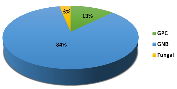
As illustrated in
Figure 1, Gram-negative bacilli (GNB) constituted
the predominant group of isolates, followed by
Gram-positive cocci (GPC). The detailed microbial
spectrum [Figure 2] demonstrated that among
Gram-negative organisms (n = 134), Acinetobacter
baumannii was the most frequently isolated
pathogen (44%), followed by Enterobacter
spp. (26.9%), Pseudomonas aeruginosa
(13.4%), Klebsiella pneumoniae (12.7%),
and Citrobacter spp. (3%). Among
Gram-positive isolates (n = 21), Staphylococcus
aureus (n = 14) predominated over Enterococcus
spp. (n = 7).
The antimicrobial
susceptibility profile of Gram-negative isolates
[Table 2] revealed widespread resistance to
cephalosporins and fluoroquinolones. Carbapenems
and aminoglycosides retained comparatively better
activity across most isolates. However, Acinetobacter
baumannii exhibited marked multidrug
resistance, with carbapenem susceptibility limited
to approximately 20%, underscoring its critical
role as a resistant nosocomial pathogen.
Among Gram-positive
organisms [Table 3], Staphylococcus aureus
demonstrated 53.8% susceptibility to cefoxitin,
indicating a substantial burden of methicillin
resistance. Moderate susceptibility was observed
to clindamycin (53.8%), while erythromycin (23.1%)
and ciprofloxacin (30.8%) showed poor activity. Enterococcus
spp. exhibited 40% susceptibility to ampicillin
and 50% high-level gentamicin sensitivity.
Notably, all Gram-positive isolates were uniformly
susceptible to vancomycin and linezolid (100%).
Comparative analysis
of MRSA and MSSA isolates (Table 4) showed that
children constituted a small proportion of both
MRSA (16.7%) and MSSA (12.5%) cases, with the
majority occurring in adults. There was no
statistically significant association between age
group and methicillin resistance (OR 1.40; 95% CI:
0.07–27.9; p = 1.00), and the wide confidence
interval reflects the limited sample size.
MRSA isolates
demonstrated lower susceptibility across multiple
antimicrobial agents compared to MSSA. Although
MSSA isolates showed higher susceptibility to
clindamycin, erythromycin, doxycycline,
ciprofloxacin, gentamicin, and cotrimoxazole,
these differences were not statistically
significant (p > 0.05). Importantly, both
MRSA and MSSA isolates remained uniformly
susceptible to vancomycin and linezolid (100%).
A particularly
noteworthy finding of the present study is the
preserved in vitro activity of colistin against
Gram-negative bacilli, including
multidrug-resistant pathogens such as Acinetobacter
baumannii and Klebsiella pneumoniae,
with the latter constituting the majority among
isolates tested for colistin susceptibility. The
observed minimum inhibitory concentration (MIC)
values ranged from 0.125 to 1 µg/mL, with no
isolate demonstrating phenotypic resistance.
The predominance of
Acinetobacter baumannii in conjunction
with its high level of carbapenem resistance
underscores its role as a critical driver of
multidrug-resistant bloodstream infections in the
ICU setting.
|
Table 1: Demographic profile of
patients with bloodstream infections in
multidisciplinary ICU
|
|
Characteristics
|
No. of Patients
|
Percentage
|
|
Age
|
0-10 years
|
06
|
3.75%
|
|
11-20 years
|
19
|
11.8%
|
|
21-30 years
|
40
|
25%
|
|
31-40 years
|
32
|
20%
|
|
41-50 years
|
16
|
10.2%
|
|
51-60 years
|
27
|
16.8%
|
|
61-70 years
|
15
|
9.3%
|
|
71-80 years
|
3
|
1.86%
|
|
81-90 years
|
2
|
1.25%
|
|
Gender
|
Male
|
72
|
45%
|
|
Female
|
88
|
55%
|
|
| Figure
1: Relative burden of major microbial
groups in bloodstream infections among
MICU patients (%) (GPC: Gram positive
cocci, GNB: Gram negative bacilli) |
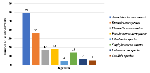
|
| Figure 2: Etiological
Spectrum of Bloodstream Infections in a
Multidisciplinary Intensive Care Unit |
|
Table 2: Antimicrobial
Susceptibility Patterns of Gram-Negative
Organisms Isolated from Blood Culture
(n=134)
|
|
Number of susceptible isolates
(%)
|
|
Antibiotics disc
|
Klebsiella pneumoniae
n=17, S (%)
|
Citrobacter species
n=04, S (%)
|
Enterobacter species
n=36, S (%)
|
Acinetobacter baumannii n=59,
S (%)
|
Pseudomonas aeruginosa
n=18, S (%)
|
|
Ciprofloxacin
|
03 (20%)
|
01 (18%)
|
07 (21%)
|
04 (8%)
|
06 (35%)
|
|
Gentamicin
|
06 (35%)
|
01 (25%)
|
11 (30%)
|
12 (20%)
|
09 (50%)
|
|
Amikacin
|
08 (50%)
|
01 (23%)
|
12 (35%)
|
15 (25%)
|
10 (55%)
|
|
Piperacillin-Tazobactam
|
05 (30%)
|
01 (24%)
|
10 (28%)
|
09 (15%)
|
08 (45%)
|
|
Ceftazidime
|
04 (25%)
|
01 (9%)
|
04 (13%)
|
06 (10%)
|
07 (40%)
|
|
Imipenem
|
08 (45%)
|
02 (47%)
|
22 (63%)
|
12 (20%)
|
11 (60%)
|
|
Meropenem
|
09 (40%)
|
02 (43%)
|
21 (60%)
|
12 (20%)
|
10 (55%)
|
|
Table 3: Antimicrobial
Susceptibility Pattern of Gram-Positive
Organisms Isolated from Blood Culture (n
= 21)
|
|
Antibiotics disc
|
Staphylococcus aureus
n=14, S (%)
|
Enterococcus spp.
n=7, S (%)
|
|
Cefoxitin
|
7 (53.8%)
|
NR
|
|
Ampicillin
|
NT
|
3 (40%)
|
|
Ciprofloxacin
|
4 (30.8%)
|
2 (30%)
|
|
Clindamycin
|
7 (53.8%)
|
NR
|
|
Erythromycin
|
3 (23.1%)
|
NR
|
|
High-level Gentamicin
|
NR
|
4 (50%)
|
|
Vancomycin
|
14 (100%)
|
7 (100%)
|
|
Linezolid
|
14 (100%)
|
7 (100%)
|
|
NR: Not Recommended, NT: Not tested
|
|
Table 4: Comparison of
Methicillin-Resistant Staphylococcus
aureus (MRSA) and
Methicillin-Susceptible Staphylococcus
aureus (MSSA) Isolates
|
|
Variable
|
MRSA
n=6 (%)
|
MSSA
n=8 (%)
|
Odds Ratio (95% CI)
|
P value,
Fisher’s exact test
|
|
Children
|
01 (16.7%)
|
01 (12.5%)
|
1.40 (0.07–27.9)
|
1.00
|
|
Adults
|
05 (83.3%)
|
07 (87.5%)
|
Reference
|
_
|
|
Antibiotics
|
N=Sensitivity (%)
|
|
Cefoxitin
|
0 (0%)
|
8 (100%)
|
NE
|
<0.001
|
|
Clindamycin
|
2 (25%)
|
5 (60%)
|
0.30 (0.03–2.85)
|
0.58
|
|
Erythromycin
|
1 (20%)
|
4 (50%)
|
0.20 (0.01–2.89)
|
0.29
|
|
Doxycycline
|
3 (51%)
|
6 (75%)
|
0.33 (0.04–2.78)
|
0.57
|
|
Ciprofloxacin
|
1 (10%)
|
5 (62.9%)
|
0.12 (0.01–1.77)
|
0.19
|
|
Gentamicin
|
2 (30.3%)
|
5 (70%)
|
0.30 (0.03–2.85)
|
0.58
|
|
Cotrimoxazole
|
3 (60%)
|
6 (80.7%)
|
0.33 (0.04–2.78)
|
0.64
|
|
Vancomycin
|
6 (100%)
|
8 (100%)
|
NA
|
_
|
|
Linezolid
|
6 (100%)
|
8 (100%)
|
NA
|
_
|
|
*NE: Not estimable, NA: Not applicable,
Reference: Reference category for
comparison.
* P-values were derived using Fisher’s
exact test. A p-value <0.05 was
considered statistically significant.
Statistically significant results are
shown in bold. Cefoxitin susceptibility
differed significantly between MRSA and
MSSA isolates (p <0.001),
confirming the discriminatory value of
methicillin resistance status.
|
Discussion
The present study
delineates the microbial spectrum and
antimicrobial resistance patterns of bloodstream
infections (BSIs) in a multidisciplinary intensive
care unit, highlighting a predominance of
Gram-negative pathogens and a high burden of
multidrug resistance.
A notable finding of
the present study was the overwhelming
predominance of Acinetobacter baumannii
among bloodstream isolates and the complete
preservation of colistin susceptibility despite
extensive resistance to multiple antimicrobial
classes. This observation provides important
contemporary data from North India, where reports
describing ICU-specific bloodstream infection
epidemiology and colistin susceptibility remain
limited.
In our cohort,
Gram-negative bacilli (GNB) accounted for the
majority of isolates, with Acinetobacter
baumannii emerging as the leading pathogen.
This finding is consistent with several studies
from Indian ICUs, where non-fermenting
Gram-negative bacilli, particularly Acinetobacter
spp., have increasingly dominated nosocomial
infections due to their environmental resilience
and remarkable ability to acquire resistance
determinants [13,14]. Similar trends have been
reported by Veeraraghavan et al. [15] and
Shrivastava et al. [16], who documented a rising
prevalence of Acinetobacter in
bloodstream infections across tertiary care
centres. Data from the Indian Council of Medical
Research (ICMR) AMR surveillance network also
highlight Acinetobacter baumannii as a
leading cause of ICU infections with high
resistance rates [17].
The high level of
resistance to cephalosporins and fluoroquinolones
observed in this study reflects the widespread
dissemination of extended-spectrum β-lactamase
(ESBL)-producing organisms and increasing
fluoroquinolone resistance. Although carbapenems
and aminoglycosides retained relatively better
activity, the susceptibility rates were
suboptimal. Of particular concern is the marked
multidrug resistance in Acinetobacter
baumannii, with carbapenem susceptibility
limited to approximately 20%. This is in
concordance with global reports of
carbapenem-resistant Acinetobacter
(CRAB), identified by the World Health
Organization as a critical priority pathogen [18].
Comparable findings have been reported in
multicentric Indian studies, which demonstrate
alarmingly high carbapenem resistance among Acinetobacter
isolates [19,20].
Among
Enterobacterales, Enterobacter spp. and
Klebsiella pneumoniae exhibited moderate
susceptibility to carbapenems, suggesting the
presence of ESBL production with evolving
carbapenem resistance. Studies by Datta et al. [21]
and Taneja et al. [22] have similarly reported
increasing resistance among Enterobacterales,
driven by ESBL and carbapenemase production. The
emergence of carbapenem-resistant Enterobacterales
(CRE) represents a major therapeutic challenge and
has been associated with poor clinical outcomes in
ICU settings [23]. Pseudomonas aeruginosa
in our study demonstrated relatively better
susceptibility to aminoglycosides and carbapenems,
although persistent resistance to fluoroquinolones
was observed, consistent with previously reported
resistance patterns [24].
Among Gram-positive
organisms, Staphylococcus aureus was the
predominant isolate, with an MRSA prevalence of
approximately 43%, aligning with reports from
Indian ICUs showing MRSA rates ranging from 30% to
50% [25,26]. MRSA isolates demonstrated reduced
susceptibility across multiple antimicrobial
classes, although statistical significance was not
achieved for most agents, likely due to the
limited sample size. Importantly, uniform
susceptibility to vancomycin and linezolid was
observed, consistent with findings from other
Indian and global studies indicating preserved
activity of these agents [27]. However, increasing
reliance on glycopeptides and oxazolidinones
raises concerns regarding the potential emergence
of resistance, including vancomycin-intermediate S.
aureus (VISA) and linezolid resistance
[28].
Enterococcus
spp., although less frequently isolated,
demonstrated reduced susceptibility to ampicillin
and variable high-level aminoglycoside resistance,
findings comparable to previous Indian studies
[29]. The absence of vancomycin resistance in the
present study is encouraging; however, the
emergence of vancomycin-resistant enterococci
(VRE) in tertiary care settings has been
increasingly reported and necessitates vigilant
surveillance [30].
The high burden of
antimicrobial resistance observed in this study
can be attributed to factors inherent to ICU
settings, including prolonged hospitalization,
invasive procedures, device-associated infections,
and extensive empirical use of broad-spectrum
antibiotics. These factors contribute to
significant selective pressure, facilitating the
emergence and dissemination of multidrug-resistant
organisms (MDROs) [31]. Similar observations have
been emphasized in global reports by the Centres
for Disease Control and Prevention (CDC) and WHO,
highlighting ICU settings as epicentres for AMR
emergence [32].
The predominance of
carbapenem-resistant Acinetobacter baumannii
observed in the present study mirrors a worrying
global trend and signals the emergence of a
potential post-carbapenem era in critical care
medicine. As resistance continues to erode the
effectiveness of conventional therapeutic options,
clinicians are increasingly compelled to rely on
last-resort agents such as colistin. The complete
susceptibility to colistin documented in this
study offers a narrow but crucial therapeutic
window; however, reports of emerging colistin
resistance worldwide warrant cautious optimism.
Sustained surveillance, rational antimicrobial
prescribing, and aggressive infection-control
measures are essential to prevent further
dissemination of multidrug-resistant organisms and
safeguard the efficacy of remaining treatment
options.
Limitations
This study is
limited by its retrospective, single-centre
design, which may affect data completeness and
generalizability. The small number of
Gram-positive isolates may have reduced the
statistical power to detect significant
differences. Additionally, molecular
characterization of resistance mechanisms and
clinical outcome analysis were not performed.
Despite these limitations, the study provides
valuable institution-specific insights into
microbial trends and antimicrobial resistance in
ICU bloodstream infections.
Conclusion
This study
highlights the emergence of carbapenem-resistant Acinetobacter
baumannii as a dominant driver of ICU
bloodstream infections and a major contributor to
the antimicrobial resistance burden. The retained
susceptibility to colistin provides a crucial
therapeutic advantage; however, reliance on
last-resort agents is unsustainable. Urgent,
coordinated efforts in surveillance, stewardship,
and infection control are essential to combat the
advancing resistance threat.
References
- Vincent JL, Rello J, Marshall J, et al.
International study of the prevalence and
outcomes of infection in intensive care units. JAMA.
2009;302(21):2323–2329.
- Singer M, Deutschman CS, Seymour CW, et al.
The Third International Consensus Definitions
for Sepsis and Septic Shock (Sepsis-3). JAMA.
2016;315(8):801–810.
- Wisplinghoff H, Bischoff T, Tallent SM, et al.
Nosocomial bloodstream infections in US
hospitals: analysis of 24,179 cases. Clin
Infect Dis. 2004;39(3):309–317.
- Timsit JF, Ruppé E, Barbier F, et al.
Bloodstream infections in critically ill
patients: epidemiology and management. Lancet
Infect Dis. 2022;22(12):e378–e390.
- Kollef MH, Zilberberg MD. Antibiotic-resistant
infections in the ICU: epidemiology and
prevention. Crit Care Clin.
2021;37(3):389–407.
- van Duin D, Paterson DL. Multidrug-resistant
bacteria in the ICU: challenges and solutions. Infect
Dis Clin North Am. 2020;34(4):709–722.
- Tamma PD, Aitken SL, Bonomo RA, et al.
Infectious Diseases Society of America guidance
on resistant Gram-negative infections. Clin
Infect Dis. 2021;72(7):e169–e183.
- O’Neill J. Tackling drug-resistant infections
globally: final report and recommendations.
London: Review on Antimicrobial Resistance;
2016.
- Donlan RM. Biofilms: microbial life on
surfaces. Emerg Infect Dis.
2002;8(9):881–890.
- O’Grady NP, Alexander M, Burns LA, et al.
Guidelines for prevention of intravascular
catheter-related infections. Clin Infect
Dis. 2011;52(9):e162–e193.
- Rhodes A, Evans LE, Alhazzani W, et al.
Surviving Sepsis Campaign: international
guidelines for management of sepsis and septic
shock. Intensive Care Med.
2017;43(3):304–377.
- World Health Organization. Global report on
the epidemiology and burden of sepsis. Geneva:
WHO; 2020.
- Diekema DJ, Hsueh PR, Mendes RE et al. The
microbiology of bloodstream infection: 20-year
trends from the SENTRY antimicrobial
surveillance program. Clin Infect Dis.
2019;69(1):e1–e9.
- Gandra S, Alvarez-Uria G, Turner P, Joshi J,
Limmathurotsakul D, van Doorn HR. Antimicrobial
resistance surveillance in low- and
middle-income countries: progress and
challenges. Lancet Infect Dis.
2020;20(9):e101–e112.
- Veeraraghavan B, Jesudason MR, Prakasah JA et
al. Antimicrobial susceptibility profiles of
Gram-negative bacteria causing infections in
India: report from ICMR network. Indian J
Med Microbiol. 2017;35(2):169–176.
- Shrivastava G, Bhatambare GS, Patel KB, Jain
S. Bacteriological profile and antibiotic
sensitivity pattern of bloodstream isolates in
ICU patients. J Clin Diagn Res.
2013;7(11):2459–2462.
- Indian Council of Medical Research (ICMR).
Antimicrobial Resistance Surveillance Network
Annual Report 2020. New Delhi: ICMR; 2020.
- World Health Organization. Global priority
list of antibiotic-resistant bacteria to guide
research, discovery, and development of new
antibiotics. Geneva: WHO; 2017.
- Manohar P, Nachimuthu R, Lopes BS. Multidrug
resistant Acinetobacter baumannii:
epidemiology, resistance mechanisms and
treatment options. Infect Drug Resist.
2020;13:2767–2785.
- Taneja N, Sharma M. Antimicrobial resistance
in the environment: the Indian scenario. Indian
J Med Res. 2019;149(2):119–128.
- Datta S, Wattal C, Goel N, Oberoi JK,
Raveendran R, Prasad KJ. A ten-year analysis of
multidrug-resistant bloodstream infections in a
tertiary care hospital. J Assoc Physicians
India. 2012;60:25–29.
- Taneja N, Kaur H. Insights into antimicrobial
resistance in India. Indian J Med Res.
2016;143(3):281–283.
- Logan LK, Weinstein RA. The epidemiology of
carbapenem-resistant Enterobacteriaceae: the
impact and evolution of a global menace. J
Infect Dis. 2017;215(Suppl 1):S28–S36.
- Bassetti M, Vena A, Croxatto A, Righi E, Guery
B. How to manage Pseudomonas aeruginosa
infections. Drugs Context.
2018;7:212527.
- Rajkumar S, Sistla S, Manoharan M, Sugumar M,
Nagasundaram N, Parija SC. Prevalence and
antimicrobial susceptibility pattern of MRSA in
ICU patients. J Lab Physicians.
2017;9(3):153–156.
- India AMR Surveillance Network.
Methicillin-resistant Staphylococcus aureus
trends in India. Indian J Med Res.
2020;151(2–3):141–149.
- Liu C, Bayer A, Cosgrove SE, Daum RS, Fridkin
SK, Gorwitz RJ, et al. Clinical practice
guidelines for MRSA infections. Clin Infect
Dis. 2011;52(3):e18–e55.
- Howden BP, Davies JK, Johnson PD, Stinear TP,
Grayson ML. Reduced vancomycin susceptibility in
Staphylococcus aureus. Clin
Microbiol Rev. 2010;23(1):99–139.
- Arias CA, Murray BE. The rise of the
Enterococcus: beyond vancomycin resistance. Nat
Rev Microbiol. 2012;10(4):266–278.
- O’Driscoll T, Crank CW. Vancomycin-resistant
enterococcal infections. Clin Microbiol Rev.
2015;28(3):747–772.
- Rosenthal VD, Al-Abdely HM, El-Kholy AA et al.
International Nosocomial Infection Control
Consortium (INICC) report, data summary of 50
countries for 2010–2015: device-associated
module. Am J Infect Control.
2016;44(12):1495–1504.
- Centers for Disease Control and Prevention
(CDC). Antibiotic resistance threats in the
United States, 2019. Atlanta: CDC; 2019.
|


