|
Introduction
Amyloidosis
comprises a heterogeneous group of disorders
characterized by extracellular deposition of
misfolded protein fibrils that exhibit Congo red
positivity with apple-green birefringence under
polarized light.[1,2] Based on distribution,
amyloidosis is classified as systemic or
localized. Localized tracheobronchial amyloidosis
(TBA) is an uncommon entity, accounting for less
than 1% of benign tracheobronchial tumors and a
very small proportion of all amyloidosis
cases.[1-3] It is most frequently associated with
AL-type (light chain) amyloid produced locally by
plasma cells.
Clinically, TBA
often presents with nonspecific symptoms such as
chronic cough, dyspnea, wheezing, recurrent
respiratory infections, or hemoptysis, and may
mimic asthma, chronic obstructive pulmonary
disease, or tracheal stenosis.[3-5] Radiological
findings are often subtle or nonspecific, making
bronchoscopy with histopathological confirmation
essential for diagnosis. Due to its rarity, there
are no standardized management guidelines, and
treatment is individualized based on symptom
severity and extent of airway involvement. We
report a case of TBA managed successfully with
bronchoscopic intervention.
Case Report
A 55-year-old male
presented to the respiratory medicine outpatient
department with complaints of chronic cough,
progressive exertional dyspnea for six months, and
hoarseness of voice. The dyspnea had gradually
worsened over time and was associated with
intermittent wheezing. He also reported occasional
episodes of scanty hemoptysis. There was no
history of smoking, pulmonary tuberculosis,
occupational exposure, connective tissue disease,
or long-term medication use. In view of the
hoarseness of voice, an otorhinolaryngology (ENT)
evaluation was sought to assess for possible vocal
cord pathology. No significant weight loss, fever,
or dysphagia was reported.
Vital signs were
stable. Respiratory system examination revealed
bilateral expiratory wheeze without stridor.
Cardiovascular, abdominal, and neurological
examinations were unremarkable.
Routine laboratory
investigations including complete blood count,
renal function tests, and liver function tests
were within normal limits. Inflammatory markers
were mildly elevated. Serum and urine protein
electrophoresis, along with serum free light chain
assay, did not reveal monoclonal gammopathy.
Autoimmune workup was negative.
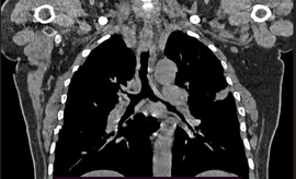
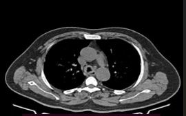
Computed tomography
(CT) of the chest demonstrated irregular
circumferential thickening of the tracheal wall
with multiple calcified nodular deposits causing
significant luminal narrowing of the trachea and
proximal main bronchi, a pattern described in
tracheobronchialpappilomatosis.[4,6] There was no
evidence of lung parenchymal mass, mediastinal
lymphadenopathy, or pleural effusion.
|
|
| Figure
1: CT chest showing tracheal wall |
Figure
2: CT chest showing tracheal wall
thickening and nodularities. |
Flexible
bronchoscopy was planned following ENT assessment.
Indirect laryngoscopy revealed nodular lesions
involving the vocal cords, raising suspicion of an
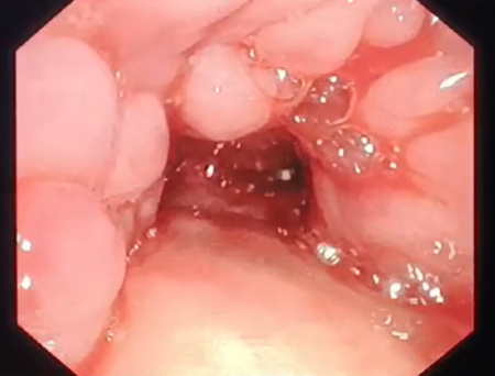
infiltrative pathology. Initial flexible
bronchoscopy demonstrated that the lesions
originated at the level of the vocal cords, with
extension into the trachea. Multiple pale-yellow,
waxy nodular lesions were seen involving the
anterior and lateral walls of the trachea,
resulting in marked luminal narrowing, which is
characteristic of tracheobronchial
papilomatosis.[3,5] The mucosa appeared firm and
brittle. Endobronchial biopsies obtained from the
accessible lesions.
|
| Figure
3: Flexible bronchoscopy revealed multiple
nodular lesions seen arising from the
trachea to both bronchial tree. |
Histopathological
examination showed subepithelial deposition of
amorphous eosinophilic material, with Congo red
positivity and apple-green birefringence under
polarized light, confirming amyloidosis.[1,2]
Congo red staining was positive, and apple-green
birefringence was demonstrated under polarized
light microscopy, confirming amyloid deposition.
Immunohistochemical analysis was consistent with
AL (lambda light chain) type amyloid. No evidence
of malignancy, granulomatous inflammation, or
vasculitis was noted.

|

|
| Figure
4: Amorphous and homogenous |
Figure
5: Congo red stain showing apple green
eosinophils deposits, birefringence under
polarized light |
Further evaluation
to exclude systemic amyloidosis included
electrocardiography, echocardiography, serum
B-type natriuretic peptide, renal evaluation, and
abdominal imaging, all of which were within normal
limits. Based on these findings, a diagnosis of
extensive tracheobronchial amyloidosis was
established.
Subsequently, the
patient underwent mechanical debulking of the
obstructing lesion followed by argon plasma
coagulation (APC) to achieve airway patency and
hemostasis. The procedure was well tolerated
without complications. Post-procedure, the patient
reported significant symptomatic improvement. In
addition, the patient was initiated on oral
colchicine at a dose of 1.5 mg/day as adjunctive
medical therapy. Follow-up bronchoscopy at three
months showed reduced lesion burden, and at six
months the patient remained asymptomatic with no
evidence of recurrence.
Discussion
Tracheobronchial
amyloidosis is a rare localized form of
amyloidosis characterized by amyloid deposition
confined to the airway walls.[1,3] The majority of
cases are of AL type, resulting from local plasma
cell dyscrasia rather than systemic disease. The
pathogenesis involves local production of
immunoglobulin light chains, which misfold and
deposit in the submucosa, leading to progressive
airway narrowing.
The clinical
presentation of TBA is often insidious and
nonspecific, resulting in frequent
misdiagnosis.[3-5] Wheezing and dyspnea may lead
to an erroneous diagnosis of asthma or chronic
obstructive pulmonary disease, while hemoptysis
and recurrent infections may raise suspicion for
malignancy or tuberculosis. Imaging findings such
as tracheal wall thickening, nodularity, and
calcification can suggest the diagnosis but are
not diagnostic.
Bronchoscopy plays a
pivotal role in both diagnosis and management of
tracheobronchial amyloidosis.[3-8] The
characteristic bronchoscopic appearance of
pale-yellow, waxy nodules with a friable or
brittle mucosa should raise suspicion of
amyloidosis. Histopathological confirmation with
Congo red staining remains the gold standard.
Importantly, exclusion of systemic amyloidosis is
mandatory, as management and prognosis differ
significantly.
There is no
definitive curative therapy for localized TBA, and
management is primarily directed toward relieving
airway obstruction and improving symptoms.[3,5,8]
Bronchoscopic interventions such as mechanical
debulking, laser therapy, argon plasma
coagulation, cryotherapy, and stent placement have
been used with varying success. Among these, argon
plasma coagulation is widely favored due to its
efficacy, precision, and favorable safety
profile.[8,9] Radiotherapy has been reported in
selected refractory cases. Systemic therapies used
for systemic amyloidosis have limited or no role
in localized disease.
The prognosis of TBA
is generally favorable compared to systemic
amyloidosis; however, recurrence is common,
necessitating long-term follow-up with periodic
bronchoscopic surveillance.[3-6] Early recognition
and timely intervention can significantly improve
quality of life and prevent life-threatening
airway compromise.
In addition to
bronchoscopic interventions, colchicine has been
used as an adjunctive medical therapy in selected
cases of localized amyloidosis, based on its
anti-inflammatory properties and ability to
inhibit amyloid fibril formation. Although
evidence supporting its use in tracheobronchial
amyloidosis is limited to case reports and small
series, colchicine may help reduce local
inflammation and slow progression when used
alongside interventional therapies.[10,11]
Conclusion
Tracheobronchial
amyloidosis is an uncommon but important cause of
unexplained airway obstruction. Its nonspecific
clinical presentation often leads to delayed
diagnosis. A high index of suspicion, combined
with radiological evaluation and confirmatory
bronchoscopic biopsy, is essential for diagnosis.
Bronchoscopic debulking with argon plasma
coagulation is an effective treatment modality
that provides symptomatic relief. Regular
follow-up is essential due to the risk of
recurrence.
References
- O'Regan A, Fenlon HM, Beamis JF, et al.
Tracheobronchial amyloidosis: the Boston
University experience from 1984 to 1999. Medicine
(Baltimore). 2000;79:69–79.
- Berk JL, O'Regan A, Skinner M. Pulmonary and
tracheobronchial amyloidosis. Semin Respir
Crit Care Med. 2002;23:155–165.
- Cordier JF, Loire R, Brune J. Amyloidosis of
the lower respiratory tract. Clin Chest Med.
2002;23:95–108.
- Capizzi SA, Betancourt SL, Martinez-Jimenez S,
Rossi SE. Tracheobronchial amyloidosis: CT
findings. J Thorac Imaging. 2010;25:W44–W47.
- Khoor A, Myers JL. Amyloidosis of the lung. Arch
Pathol Lab Med. 2015;139:1008–1013.
- Milani P, Basset M, Russo F, et al. The lung
in amyloidosis. Eur Respir Rev.
2017;26:170046.
- Shah PL, Gilligan D, Hawkins T, et al.
Endobronchial amyloidosis treated with argon
plasma coagulation. Thorax.
2001;56:328–330.
- Gillmore JD, Hawkins PN. Amyloidosis and the
respiratory tract. Thorax. 1999;54:444–451.
- Sipe JD, Benson MD, Buxbaum JN, et al. Amyloid
fibril proteins and amyloidosis: nomenclature
2010. Amyloid. 2010;17:101–104.
- Utz JP, Swensen SJ, Gertz MA. Pulmonary
amyloidosis: the Mayo Clinic experience from
1980 to 1993. Ann Intern Med.
1996;124:407–413.
- Lachmann HJ, Goodman HJ, Gilbertson JA, et al.
Natural history and outcome in systemic AA
amyloidosis. N Engl J Med. 2007;356:2361–2371.
|


