|
Introduction
Cervical
lymphadenopathy is a common manifestation of
underlying pathologies related to the head and
neck, with a wide range of differential diagnoses.
Non-neoplastic conditions more frequently affect
lymph nodes compared to neoplastic processes.[1,2]
Among neoplastic lesions, lymph node metastasis is
most prevalent, with the primary site often being
of squamous cell origin, as noted in few studies.
Recent studies highlight the role of Human
Papilloma Virus(HPV) in causation of head and neck
squamous cell carcinoma.[3,4]
Fine-needle
aspiration cytology (FNAC) is generally the
initial assessment tool for cervical
lymphadenopathy. However, cervical lymph node
metastasis of squamous cell carcinoma (SCC) poses
diagnostic challenge on FNAC when the tumour is
well differentiated and lack nuclear atypia.[5]
Many times, secondary changes like necrosis,
abscess formation or cystic change may occur in
the metastatic lymph node making the diagnosis on
cytology a difficult task. [6, 7] Also
many benign conditions like infected keratinous
cyst or squamous metaplasia mimic well
differentiated SCC, adding to the diagnostic
dilemma. Repeat aspirate or guided FNAC may demand
time, cost and expertise.
Recently, Hara H et
al introduced the Five Parameter System (FPS)
which is useful in differentiating oral SCC from
in situ carcinoma on cytology. [8] This system
focuses solely on assessing keratinized cellular
findings for cytological examination of oral
well-differentiated squamous cell carcinoma (SCC)
and carcinoma in situ (CIS) specimens. These
specimens can sometimes lack nuclear atypia,
making them difficult to categorize accurately
within The Bethesda System (TBS). [8]
Furthermore, same
authors tested the utility of this system in
differentiating oral SCC from in situ carcinoma
when nuclear atypia is present and concluded that
recognition of the following five parameters which
are concentric arrangement of orangeophilic cells,
large number of cells, bizarre-shaped cells
without nuclear atypia, keratoglobules and uneven
filamentous cytoplasm are useful for the
cytological diagnosis of oral SCC. [9]
Hence, the present
study was planned to determine the usefulness of
five parameter system in diagnosing metastatic
deposits of SCC to lymph node with secondary
changes.
Objectives:
To determine the
utility of five parameter system in diagnosing
metastatic SCC showing secondary changes like
cystic degeneration or abscess formation
Material and Methods
The present study
was conducted at a tertiary care rural hospital.
Institutional Ethics Committee clearance (IEC) was
obtained from IEC-AIMS approval number being
42/2018 dated 10th January 2018. This
is a cross sectional study, conducted for a period
of 04 years, from 2018 to 2022. The study
population composed of 70 cases and 45 controls.
Sampling technique used was purposive sampling.
The study population composed of all the patients
who had cervical lymphadenopathy who underwent the
procedure of fine needle aspiration in the
department of pathology in the institute with a
cytological diagnosis of metastatic squamous cell
carcinoma (SCC) with cystic change or abscess
formation, with biopsy confirmation wherever
available. Forty five cases of inflamed keratinous
cyst diagnosed on FNAC were taken as controls in
the study. Written informed consent was obtained
before the study from all the study participants
after providing adequate information about the
study. The demographics of study population were
recorded from case records for all the patients.
The FNAC slides from
all 634 patients presenting with cervical
lymphadenopathy were reviewed by two pathologists,
each with over ten years of cytology experience.
For this study, all the cases in whom FNAC
diagnosis of metastatic SCC to cervical lymph
nodes with inflammation/cystic change/abscess
formation and had histological confirmation
wherever available were selected. In deep seated
lymph nodes where biopsy was not feasible, however
which had a clinically confirmed primary,
radiology was used as a supportive tool for
delineating metastatic lymph nodes deposits which
were then confirmed by FNAC. The procedures
followed in the present study adhered to the
ethical guidelines of the Declaration of Helsinki.
Inclusion
criteria:
Cases: FNAC cases of
metastatic SCC to cervical lymph nodes with cystic
change or abscess formation, with biopsy
confirmation wherever available. Amongst cases,
those smears which showed presence of >30
diagnosable epithelial cells were included.
Controls: 45 cases
of inflamed keratinous cyst diagnosed on cytology
were taken as controls. Among controls those
smears which has >200 squamous epithelial cells
smeared on slide were included.
All smears from both
cases and controls were evaluated for the Five
Parameter System(FPS) focusing on cytoplasmic
features which included concentric arrangement of
orangeophilic cells, large number of orangeophilic
cells, bizarre shaped orangeophilic cells,
keratoglobules and dyskeratosis. (Figure 1 to 4)

|
| Figure
1: A and B: FNAC smears showing concentric
arrangement of squamous cells in cases
i.e. metastatic deposits of squamous cell
carcinoma. (Hematoxylin and Eosin, x 200)
|

|
| Figure 2: FNAC smears
showing orangeophilic cells in cases i.e.,
metastatic deposits of squamous cell
carcinoma. (Hematoxylin and Eosin, x 200) |

|
| Figure 3: A and B: FNAC
smears showing bizarre shared
orangeophilic cells in cases i.e.,
metastatic deposits of squamous cell
carcinoma. (Hematoxylin and Eosin, x 200) |

|
| Figure 4: FNAC smears
showing keratoglobules in cases i.e.,
metastatic deposits of squamous cell
carcinoma. (Hematoxylin and Eosin, x 200) |
Statistical
analysis:
Descriptive
statistics was used like mean and standard
deviation, percentages and proportions. Difference
between two groups was analysed using unpaired t
test with p<0.05 set as statistical
significance.
Results
The total number of
FNACs done on patients with cervical
lymphadenopathy at the study institute, during the
study period was 634 which constituted 8.05% of
the total FNACs done. A total of 70 cases and 45
controls were included in this study. Of the total
70 cases, 56 cases of SCC were well-differentiated
SCC and 14 cases were poorly differentiated SCC.
Demographic characteristics of study population is
depicted in Table 1.
|
Table 1: Demographic
characteristics of the study population
|
|
Variables
|
Control
|
Cases
|
|
Age (years±SD)
|
38±1
|
62±2
|
|
Female to male ratio
|
1:1.5
|
1:2
|
|
Site of Primary in descending order
|
Skin of Head and neck region (60.5%),
extremities (20%) and back (19.5%)
|
Lip (30%)
Buccal mucosa (20%)
Pyriform fossa (20%)
Oesophagus (20%)
Tonsil (05%)
Skin (05%)
|
The FNAC smears of
all the cases and controls were evaluated using
the five parameter system, Among the five
parameters assessed, in the cases; the most
frequently observed feature was large number of
orangeophilic cells, seen in 58 cases (82.8%).
This was followed by uneven filamentous cytoplasm
in 50 cases (71.4%), bizarre-shaped orangeophilic
cells in 46 cases (65.7%), a concentric
arrangement of cells in 6 cases (8.57%), and
keratoglobules in 2 cases (2.85%). In contrast,
none of these characteristics were present in the
control group (Table 2).
|
Table 2: Comparison of 5
parameter system between SCC and
inflamed keratinous cyst
|
|
Sl No
|
Parameter assessed
|
SCC (70)
|
Inflamed keratinous cyst (40)
Inflamed keratinous cyst (40)
|
|
1
|
Concentric arrangement
|
06 (8.57%)
|
0%
|
|
2
|
Large numbers of orangeophilic cells.
|
58 (82.8%)
|
0%
|
|
3
|
Bizarre-shaped orangeophilic cells
|
46 (65.7%)
|
0%
|
|
4
|
Keratoglobules
|
02 (2.85%)
|
0%
|
|
5
|
Uneven filamentous cytoplasm
|
50 (71.4%)
|
0%
|
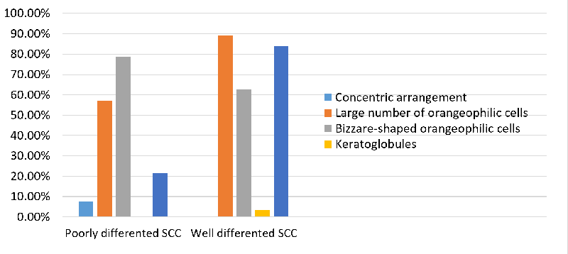
Out of the 70 cases
of metastatic SCC, 56 were well-differentiated and
14 were poorly differentiated. (Table 3, Figure 5)
Concentric cell arrangements were observed in 5
cases (8.92%) of well-differentiated SCC and in 1
case (7.8%) of poorly differentiated SCC. A large
number of orangeophilic cells appeared in 50 cases
(89.2%) of well-differentiated SCC and 8 cases
(57.14%) of poorly differentiated SCC.
Bizarre-shaped orangeophilic cells were found in
35 cases (62.5%) of well-differentiated SCC and in
11 cases (78.57%) of poorly differentiated SCC.
Keratoglobules were present in 2 cases (3.57%) of
well-differentiated SCC, while none were seen in
poorly differentiated SCC. Uneven filamentous
cytoplasm was noted in 47 cases (83.92%) of
well-differentiated SCC and 3 cases (21.42%) of
poorly differentiated SCC (Table 3 and Figure 5).
Over all there was statistically significant
difference between the frequency of all the 5
parameters among cases and control (unpaired t
test, p<0.05)
|
Table 3: Comparison of 5
parameter system between
well-differentiated and poorly
differentiated SCC
|
|
SL No
|
Parameter assessed
|
Well differentiated SCC (N-56)
|
Poorly differentiated SCC (N-14)
|
|
1
|
Concentric arrangement
|
05 (8.92%)
|
01(7.8%)
|
|
2
|
Large numbers of orangeophilic cells.
|
50(89.2%)
|
08(57.14%)
|
|
3
|
Bizarre-shaped orangeophilic cells
|
35(62.5%)
|
11(78.57%)
|
|
4
|
Keratoglobules
|
2(3.57%)
|
00
|
|
5
|
Uneven filamentous cytoplasm
|
47 (83.92%)
|
03(21.42%)
|
|
Unpaired t test, p<0.05
|
|
| Figure
5: Graphical representation of comparison
of 5 parameter system between well
differentiated SCC and poorly
differentiated SCC |
Discussion
Finding of
keratinized squamous cells in a lymph node is a
strong pointer to metastasis of SCC, however, in
the presence of cystic degeneration or abscess
formation, which is commonly seen in cervical
metastasis of SCC, FNAC smears demonstrate
necrosis and acute suppuration, whereas atypical
cells are very scanty. Moreover, the degree of
atypia is either less or may be attributed to the
reactive changes, leading to false negative
interpretation. Though lymph nodes and keratinous
cyst can be differentiated clinically, this
differentiation is not straightforward and
ambiguities do occur. For example, the cystic
lesions, when inflamed or rupture, develop pain,
erythema and adhesions which may mimic inflamed
lymph nodes. Similarly, the lymph nodes when they
undergo suppuration or cystic changes may appear
as soft fluctuant mass, mimicking cystic lesions.
Moreover, at cytology, the inflammatory atypia and
metaplastic changes in an inflamed keratinous cyst
is a common pitfall for a false positive diagnosis
of SCC, especially if the diagnosis is based
solely on keratin presence. The five-parameter
system is an indispensable adjunct tool in these
diagnostically ambiguous cases which takes into
consideration the more reproducible cytoplasmic
keratinization-based features rather than nuclear
characteristics alone. Hence, the present study
emphasizes the usefulness of FPS in
differentiating metastatic SCC from its benign
mimickers, especially in cytologically equivocal
or paucicellular aspirates.
Lymph node
metastasis in cervical region have primary tumours
more commonly in the oropharynx. tonsil and base
of tongue. HPV is a well-established risk factor
for the squamous cell carcinoma in these regions
with significant prognostic and therapeutic
implications. Imaging modalities like Computed
Tomography, Magnetic Resonance Imaging along with
FNAC plays a crucial role in the diagnostic
workup. [5, 6]
False negative rate
in metastatic squamous cell carcinoma is a cause
of concern since most are well differentiated and
lack nuclear atypia. Hara et al (2017) observed in
their study, that the five parameter system was
successful in discriminating oral SCC.[9] These
five parameter system (FPS) is based on
cytoplasmic features and hence was proposed to be
useful in cases which lacked nuclear atypia.
Recently, Hara et al (2022), applied this FPS in
oral SCC with apparent nuclear atypia. [8] (Table
4)
|
Table 4: Comparison of 5
parameter system finding in the present
study with that reported in the
literature
|
|
Author
|
Cases
|
Concentric arrangement
|
Large numbers of orangeophilic
cells
|
Bizarre-shaped orangeophilic
cells
|
Keratoglobules
|
Dyskeratosis
|
|
Hara et al (2017)
|
Cases
Oral SCC-12
|
75%
|
100%
|
83.3
|
100
|
100
|
|
Carcinoma in situ - 04
|
0%
|
75%
|
75
|
100
|
100
|
|
Control-Non neoplastic lesions of oral
mucosa -04
|
0%
|
0%
|
0%
|
0%
|
0%
|
|
Present study
|
Cases- Metastatic deposits of SCC with
inflammation and cystic change
Well differentiated-56
|
8.92%
|
89.2%
|
62.5%
|
3.57%
|
83.92%
|
|
Cases- Metastatic deposits of SCC with
inflammation and cystic change
Poorly differentiated-14
|
7.8%
|
57.14%
|
78.57%
|
0%
|
21.42%
|
|
Control-
|
0%
|
0%
|
0%
|
0%
|
0%
|
In the present
study, we attempted to apply the FPS in metastatic
SCC, especially in cases which showed inflammation
and cystic change.
The current study
investigated cytomorphological characteristics of
metastatic squamous cell carcinoma (SCC) in
patients with cervical lymphadenopathy, focusing
on differences between well-differentiated and
poorly differentiated cases. A comparative
analysis of these cases with controls revealed
distinct cytological features that can aid in the
identification and differentiation of metastatic
SCC.
A previous study by
Hara et al (2017) has showed high frequency of
orangeophilic cells (100%) in SCC cases. [9]
Similar observations were made in the present
study. The high frequency of orangeophilic cells,
observed in 89.2% of well-differentiated and
57.14% of poorly differentiated cases in our
study, underscores their diagnostic significance
in SCC. This characteristic was the most
frequently observed feature among the five
parameters assessed, seen in 82.8% of all cases,
yet absent in controls. This finding aligns with
previous studies that highlight the presence of
orangeophilic cells as a hallmark of SCC due to
the presence of keratinized cytoplasm, a key
feature in well-differentiated tumors. [8] The
higher incidence of orangeophilic cells in
well-differentiated cases may reflect the greater
degree of keratinization compared to poorly
differentiated SCC.
The presence of
bizarre-shaped orangeophilic cells, observed in
62.5% of well-differentiated and 78.57% of poorly
differentiated SCC, further supports the
cytomorphological variations within SCC subtypes.
This parameter was seen in 65.7% of all SCC cases,
indicating its diagnostic utility in identifying
malignant features within SCC, particularly in
distinguishing between SCC subtypes based on
cellular morphology. The higher frequency of these
cells in poorly differentiated cases may be
attributed to increased cellular atypia, a
hallmark of lower differentiation grades in SCC.
Similar observations of increased bizarre-shaped
orangeophilic cells (83.3%) were documented by
Hara et al. [8]
The concentric
arrangement of cells, although less frequent, was
observed in 8.92% of well-differentiated and 7.8%
of poorly differentiated SCC cases. While this
feature is not as prominent as orangeophilic or
bizarre-shaped cells, its presence still
contributes to the identification of SCC, as it
reflects the organized, albeit disrupted, cellular
architecture commonly seen in neoplastic lesions.
Hara et al has documented the concentric
arrangement of cells in 75% of the cases of SCC,
which is high compared to our study. [8]
Keratoglobules were
documented in all the cases of SCC (100%) by Hara
et al. [8] in the present study, Keratoglobules,
noted in only 3.57% of well-differentiated cases
and absent in poorly differentiated cases, were
the least common feature identified. Their limited
occurrence suggests that keratoglobules may not be
a reliable diagnostic marker in SCC, particularly
for poorly differentiated cases. In contrast to
Hara et al. who included FNAC of primary SCC in
their study, the present study included FNAC from
metastatic SCC deposits in lymph nodes. Most of
these cases harbour necrosis, inflammation or
demonstrate cystic degeneration, wherein
keratinization may be incomplete or disrupted.
Formation of concentric cell arrangements and the
presence of keratoglobules reflect mature
keratinization; and the same is reduced in
metastasis, especially in the presence of
secondary changes. Likewise, the variation in
cellularity and tumour differentiation in primary
and metastatic process may have contributed to the
decreased prominence of keratinized structures.The
absence of keratoglobules in poorly differentiated
cases may correlate with reduced keratinization, a
characteristic of lower differentiation. While
their lower frequency may limit their utility as
primary diagnostic criteria, these features, when
present, could serve as ancillary markers, further
substantiating the diagnosis of SCC.
Uneven filamentous
cytoplasm, observed in 83.92% of
well-differentiated and 21.42% of poorly
differentiated cases, ranked second in frequency
among the parameters. This characteristic, present
in 71.4% of all SCC cases, highlights its
relevance as a diagnostic feature, especially for
well-differentiated tumors. This result is in
concordance with the study by Hara et al, in which
the uneven filamentous nature of the cytoplasm has
been documented in 100% the cases of SCC. [8] The
uneven filamentous nature of the cytoplasm may
indicate the presence of keratinized strands, a
prominent feature of well-differentiated SCC,
which is typically reduced or absent in poorly
differentiated variants.
The complete absence
of these cytological features in the control group
underscores their potential specificity. This
specificity enhances the diagnostic accuracy of
FNAC in these cases, offering a rapid, minimally
invasive, and cost-effective approach to the
diagnosis of metastatic SCC.
Limitations
Limited sample size
and single institution study is the major
limitation of the present study. In the study
conducted by Hara et al, authors included primary
SCC cases, however the present study had strict
inclusion criteria and specifically focused on the
metastatic deposits of SCC with cystic
degeneration which is less common and is a more
problematic area, especially in the context of the
overlap with benign cystic keratinous lesions.
Hence, the present study cohort reflects real
incidence patterns over 4 years. Moreover, the
present study is one among the few studies
evaluating FPS in metastatic SCC. Further studies
with large sample size will be imperative to test
the diagnostic value of FPS
Conclusion
Five parameter
system can serve as a powerful tool in FNAC
diagnosis of metastatic deposits of SCC in
challenging situations such as presence of cystic
change and abscess formation. The distinct
cytomorphological features, such as the presence
of orangeophilic cells, bizarre-shaped cells, and
uneven filamentous cytoplasm, provide valuable
diagnostic markers for metastatic SCC, and helps
in distinguishing between well-differentiated and
poorly differentiated forms
References
- Chabra S, Mohan H, Bal A. A Retrospective
Histological Evaluation of Non-neoplastic
Superficial Lymphadenopathy. Int J Internal
Med. 2005;6(1):1-5.
- Kanhere S, Seurange S, Khan SS et al.
Evaluation of FNAC in lymphadenopathy. Ind J
Surg. 1994;56:169-74.
- Dasari P, Varani S, Pattnayak S, Nagababu,
Nandini. Cervical Lymphadenopathy: A Prospective
Study in Rajiv Gandhi Institute of Medical
Sciences, Srikakulam, Andhra Pradesh. Int J
Sci Stud. 2016;4(5):233-8.
- Prasad S, Mohan N. Efficacy of aspiration
cytology in suspected metastatic neck lymph
nodes. Int J Med Sci Public Health.
2014:3;46-8.
- Pindborg JJ, Reichart PA, Smith CJ, van der
Waal I. Histological typing of cancer and
precancer of the oral mucosa. WHO Classification
of Tumours. 2nd ed. Berlin: Springer; 1997. p.
24-26.
- Konar K, Ghosh S, Ghosh T, Bhattacharya S,
Sanyal S. Pitfalls in the cytodiagnosis of
metastatic squamous cell carcinoma in the head
and neck: A retrospective study. J Cytol.
2008;25(4):119-22.
- Sahni S, S VS, Krishnappa A. Diagnostic
efficacy of fine needle aspiration cytology in
cystic lesions of head and neck region-A single
experience at tertiary health care centre. National
Journal of Laboratory Medicine.
2017:6(2):1-7
- Hara H, Misawa T, Ishii E et al. Usefulness of
Five-Parameter System Reconfirmed for
Cytopathology of Oral Squamous Cell Carcinoma
regardless of Differentiation Degree. Acta
Cytol. 2022;66(3):216-27.
- Hara H, Misawa T, Ishii E et al. Differential
diagnosis of well-differentiated squamous cell
carcinoma from non-neoplastic oral mucosal
lesions: New cytopathologic evaluation method
dependent on keratinization-related parameters
but not nuclear atypism. Diagn Cytopathol.
2017;45(5):406-17.
|


