|
Introduction
The
larynx is an endogenous anatomical structure that
serves a multifaceted purpose, including
safeguarding, breathing, and vocalisation. In
order to perform its intended role, it is
necessary for the larynx to possess adaptability
and flexibility. The proper configuration of the
vocal cord's structure is crucial for speech
production. This structure comprises the
epithelium and lamina propria of a vocal ligament,
with particular emphasis on the superficial layer,
also known as Reinke's space. This layer is of
utmost importance as it represents a potential
space with minimal subepithelial connective
tissue, as documented in literature [1].
Laryngeal lesions
encompass a range of conditions, including
non-cancerous growths on the vocal cords, as well
as potentially cancerous growths that may be
either precursors to malignancy or fully malignant
in nature. The prevalent benign laryngeal lesions
are vocal nodules and polyps. These are typically
identified through patient history, clinical
complaints, and visual examination techniques,
such as indirect laryngoscopy with rigid or
flexible fibre optic scope and stroboscopy [2].
Hoarseness is a prevalent laryngeal symptom that
prompts patients to seek treatment in the field of
otolaryngology. This condition can be attributed
to a range of potential disorders.
Currently, the
diagnostic process for laryngeal diseases in
clinical settings is intricate and relies on the
assessment of patients' symptoms, medical history,
and results from instrumental and histological
examinations. Over the past 20 years, numerous
imaging techniques have been developed to examine
the larynx and acquire precise measurements of
voice quality [3]. Recent research has highlighted
the significance of the clinico-histological
correlation in laryngeal pathologies. Therefore,
it is imperative to conduct a histological
examination in order to ascertain the
characteristics of laryngeal lesions and determine
subsequent management strategies.
Materials and Methods
The present
investigation was carried out at the Department of
Otorhinolaryngology situated in Chettinad Hospital
and Research Institute. The study was conducted in
accordance with the guidelines set forth by the
Institutional Human Ethical Committee and spanned
from February 2022 to April 2023. The research
encompassed a cohort of 80 specimens comprising
individuals who received surgical intervention for
laryngeal lesions. Following the acquisition of
informed consent from the participants, a
histopathological analysis was conducted on each
sample. This was achieved by initially fixing the
samples with buffered formalin (10%). Hematoxylin
and eosin stains were utilised for the
examination, and for certain cases, an
immunohistochemical study was conducted. The
pertinent clinical information was extracted from
the case files. The slides were gathered and
subjected to analysis to investigate diverse
clinicopathological manifestations of laryngeal
lesions.
Results
The findings of our
study indicate a higher prevalence of the
condition among males as compared to females, with
a male to female ratio of 1.17:1. Regarding the
patients' professions, 27 individuals identified
as housewives, 7 individuals identified as
teachers, and 4 individuals identified as singers.
Furthermore, the study revealed that 38 (47.5%) of
the patients were identified as smokers, while the
remaining 42 (52.5%) were classified as
non-smokers. The prevalent grievance among 72
patients (90%) is the manifestation of hoarseness
in their vocal quality. The study revealed that a
significant proportion of patients experienced
various complaints, such as Dysphagia in 36
patients (45%), Dyspnoea in 25 patients (31.25%),
Odynophagia in 10 patients (12.05%), Aspiration in
2 patients (2.5%), and hemoptysis in 1 patient
(1.02%).
Out of a cohort of
80 patients, 46.25% (n=37) presented with
non-neoplastic lesions, while 53.75% (n=43) were
diagnosed with neoplastic lesions. The study
revealed that among the 37 non-neoplastic lesions,
vocal cord polyp was the most frequently occurring
lesion, with a prevalence of 48.64% among the
patients (as presented in Table 1). Similarly,
among the 43 neoplastic lesions, squamous cell
carcinoma was found to be the most prevalent
lesion, with a prevalence of 74.41% among the
patients (as presented in Table 2).
|
Table 1: Distribution of
non-neoplastic lesions
|
|
Lesions
|
Number of cases
|
Percentage
|
|
Vocal cord polyp
|
18
|
48.64%
|
|
Vocal cord nodule
|
13
|
35.13%
|
|
Keratosis
|
3
|
8.10%
|
|
Vocal cord cyst
|
2
|
5.40%
|
|
Laryngeal cyst
|
1
|
2.70%
|
|
Table 2. Distribution of
Neoplastic Lesions
|
|
Neoplastic
|
Lesions
|
Number of cases
|
Percentage
|
|
Benign
8 (18.60%)
|
Papilloma
Granuloma
Hemangioma
Pleomorphic adenoma
|
5
1
1
1
|
11.62%
2.32%
2.32%
2.32%
|
|
Malignant
35 (81.39%)
|
Squamous cell carcinoma
Adenocarcinoma
|
32
3
|
74.41%
6.97%
|
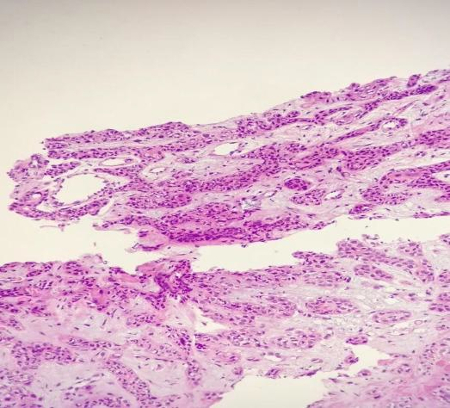
A polyp of the vocal
cord is a pedunculated mass that is round and
circumscribed, and it develops along the
membranous portion of the vocal cord. The
histological presentation of the subject matter
may manifest in gelatinous, telangiectatic, or
hyaline form. The lamina propria exhibits
alterations in its vasculature, deposition of
amorphous material, and evidence of prior
hemorrhagic events, as depicted in Figure 1.

|
| Fig.
1: Vocal cord polyp |
Vocal cord nodules
are non-malignant, greyish-white, and firm
protrusions that are located along the unattached
edge of the vocal cords. It typically manifests at
the intersection of the anterior one-third and
posterior two-thirds. Females are more frequently
impacted by voice misuse and improper vocal
technique. The histopathological examination
reveals the presence of submucosal edema and
haemorrhage, which subsequently undergo
hyalinization and fibrosis. The formation of a
nodule can be observed as a result of hyperplasia
of the overlying epithelium (refer to Fig. 2).

|
| Fig
2: Vocal cord nodule |
Keratosis refers to
the presence of a white plaque or warty growth on
the upper surface of one or both vocal cords,
which is attributed to prolonged exposure to
irritants. The aforementioned is a lesion that has
the potential to develop into cancer. Epithelial
hyperplasia will be observed at a microscopic
level, as depicted in Figure 3.

|
| Fig.
3: Keratosis |
Vocal cord cysts are
non-malignant growths that manifest as a sac-like
structure enclosed within the membranous vocal
cords. From a histological standpoint, it can be
classified into two distinct types: Vocal cord
cysts can be classified into two types:
subepithelial vocal cord cysts and ligament vocal
cord cysts. The former occurs in the superficial
lamina propria, while the latter occurs in the
deep layers of the propria. The lining of the cyst
wall comprises of either squamous epithelial cells
or glandular cells, as depicted in Figure 4.

|
| Fig
4: Vocal cord cyst |
A benign lesion
known as the laryngeal cyst is known to occur
within the mucosal layer of the larynx, with a
higher incidence in the supraglottic region. This
occurrence can result in respiratory distress.
From a histological perspective, the entity in
question can be classified into five distinct
types, namely Ductal (constituting 75% of cases),
Saccular (accounting for 24% of cases), and
several other subtypes including Laryngocele,
Oncocytic cyst, and Tonsillar cyst. The lining of
the cyst wall is composed of glandular cells, as
depicted in Figure 5.

|
| Fig.
5: Laryngeal cyst |
Papilloma is a
pathological state that arises due to mucosal
infection by human papillomavirus and is
considered to be a precursor to malignancy. The
male population exhibits a higher incidence rate
of the aforementioned condition. The histological
features of the condition are distinguished by the
presence of papillary proliferation of stratified
squamous epithelium, which may exhibit varying
degrees of hyperkeratosis or parakeratosis.
Granulations of the
vocal cords are infrequent and may present as
either unilateral or bilateral rounded lesions of
diverse hues, featuring a pedicle and a surface
that may be either smooth or uneven. At the
microscopic level, the observed features include
an undamaged or ulcerated squamous epithelium,
accompanied by the growth of fibrous tissue and
highly vascularized granulation tissue.
Additionally, there is infiltration of lymphocytes
and plasma cells, as depicted in Figure 6.

|
| Fig.
6: Granulation tissue |
Laryngeal
hemangiomas are vascular tumours that exhibit slow
progression and are frequently detected in
paediatric patients. The pathological analysis
reveals the presence of hyperplasia of blood
vessels and hemangiectasis beneath the squamous
mucosa, accompanied by lymphocyte infiltration
surrounding the vessel.
Pleomorphic adenomas
are infrequent pathological occurrences observed
within the larynx. Typically, the tumour is
characterised by a slow growth rate, solitary
appearance, and absence of pain. Pleomorphic
adenomas are typified by the presence of
epithelial tissue intermingled with tissues
exhibiting myxoid, mucoid, or chondroid
morphologies. The pleomorphic adenoma of the
larynx may exhibit similarities to aggressive
epithelial tumours in terms of its histological
features, owing to its high cellularity and
absence of a stromal component, as depicted in
Figure 7.

|
| Fig.
7: Pleomorphic adenoma |
Squamous cell
carcinoma represents the most prevalent form of
malignant neoplasm. The examination conducted at a
microscopic level reveals the presence of tumour
cells that are grouped together in masses or
nests, exhibiting squamous differentiation. These
groups are characterised by the presence of
extracellular keratin pearls, intracellular
keratin, and intercellular bridges, as depicted in
Figure 8.
|
| Fig.
8: Squamous cell carcinoma |
Laryngeal
malignancies comprise a minute fraction of
adenocarcinomas, constituting less than one
percent of the total cases. The tumour tissue
exhibited a lobulated morphology and consisted of
polygonal epithelial cells, as observed through
histological analysis. The cellular entities
coalesce into cohesive clusters and configurations
reminiscent of acinar formations.
Discussion
Laryngeal neoplasms
are typically identified at an early stage as a
result of dysphonia, which is characterised by
alterations in vocal quality. The most frequently
reported symptom in our investigation was
hoarseness of voice, with a prevalence of 90%. The
prevalence of benign tumours was highest during
the third decade of life, while malignant tumours
were more frequently observed among individuals
aged 61 to 70 years. According to Shirley D and
colleagues, there is a positive correlation
between age and the incidence of malignant
tumours, with the mean age of diagnosis being 66
years [4].
The present study
revealed a higher incidence of both benign and
malignant lesions in males as compared to females,
with a male to female ratio of 1.17:1. These
findings are consistent with those reported by
Wani et al. The higher prevalence of voice
disorders in males may be linked to factors such
as occupational hazards, vocal misuse, smoking,
and alcohol consumption, as reported in previous
studies [5].
Approximately 46.25%
of cases were comprised of non-neoplastic lesions,
with vocal polyps being the most frequently
occurring type. The vocal cord polyp is a
discrete, pedunculated lesion that develops along
the membranous portion of the vocal cord. The
histological presentation of the specimen may
exhibit characteristics of either the gelatinous,
telangiectatic, or hyaline type. The present study
revealed a prevalence of 22.5% of vocal polyp
cases, which is higher than the prevalence
reported by Chopra et al. (16%) and lower than the
prevalence reported by Kavitha Y et al. (31.4%) in
their respective studies [6,7].
The second most
frequent non-neoplastic lesion observed was vocal
cord nodule, which accounted for 13 cases
(16.25%). Varalakshmi KP and colleagues reported a
prevalence of 8.4% in their study [8]. The study
identified additional non-neoplastic lesions,
including keratosis, vocal cord cyst, and
laryngeal cyst.
The prevalence of
neoplastic lesions was found to be 53.75%, with
10% of cases being benign tumours and 43.75% being
malignant tumours. Papilloma was found to be the
most prevalent type among the benign neoplastic
lesions. Papilloma is a pathological state that
arises due to mucosal infection by human
papillomavirus and is considered to be a
precancerous condition. The histological features
of this condition are marked by the papillary
proliferation of stratified squamous epithelium,
which exhibits varying degrees of hyperkeratosis
or parakeratosis. The study observed a prevalence
of 6.25%, which is consistent with the findings of
Chavan SS et al. [9] and Ritu Bhagatet al., who
reported a prevalence of 6% and 6.7%,
respectively. Our study observed one instance of
hemangioma cases, which accounts for 1.25% of the
total cases. This finding is consistent with the
results reported by Varalakshmi KP et al and Ritu
Bhagat et al, who also observed a similar
incidence rate of 1.1%.
The most prevalent
malignant neoplasm observed was squamous cell
carcinoma, accounting for 40% of cases with a
total of 32 instances. In their respective
studies, Varalakshmi KP et al, Ritu Bhagat et al,
and Kavitha Y et al documented prevalence rates of
41.1%, 52.2%, and 36.4% for the condition under
investigation. Squamous cell carcinoma frequently
originates in a context of mucosal squamous
dysplasia or carcinoma in situ. The presentation
typically involves the infiltration of the
laryngeal stroma by atypical cells in the form of
islands, tongues, and clusters. The study reported
a prevalence of 3 cases (3.75%) of adenocarcinoma
of the larynx, which is higher than the prevalence
reported by Varalakshmi KP et al and Ritu Bhagat
et al (1.1%) in their respective studies [10].
Conclusion
The manifestation of
laryngeal tumours can exhibit a range of severity,
from minor changes in vocal quality to critical
respiratory compromise [11]. It is imperative to
employ all feasible diagnostic techniques to
expedite the identification of malignant tumours,
given their significantly elevated incidence. The
timely detection of a lesion through endoscopic
examination and subsequent biopsy can result in
successful treatment and significantly enhance the
patient's prognosis [12]. Individuals who are
considered high-risk, such as elderly males with a
smoking history, should be thoroughly assessed
with a heightened level of suspicion for the
presence of malignant lesions if they exhibit any
of these symptoms [13].
References
- Suárez-Quintanilla J, Cabrera AF, Sharma S.
Anatomy, head and neck, larynx. StatPearls
[Internet]. 2020 Sep 8.
- Malik P, Yadav SP, Sen R, Gupta P, Singh J,
Singla A, Vashisht S. The clinicopathological
study of benign lesions of vocal cords. Indian
Journal of Otolaryngology and Head and Neck
Surgery. 2019 Oct;71(1):212-20.
- Verikas A, Uloza V, Bacauskiene M, Gelzinis A,
Kelertas E. Advances in laryngeal imaging. European
Archives of Oto-rhino-laryngology. 2009
Oct;266(10): 1509-20.
- Shirley D. Cartilaginous lesions of the
larynx. Grand rounds archives BCM. Bobby R
Alford Department of Otolaryngology - Head and
Neck Surgery. 1997
- Wani AA, Rehman A, Hamid S, Akhter M, Baseena
S. Benign mucosal fold lesion as a cause of
hoarseness of voice. A Clinical study. Otolaryngology.
2012;2(3):120.
- Chopra H, Kapoor M. Study of benign glottis
lesions undergoing microlaryngealsurgery. Indian
J Otolaryngol Head Neck Surg.
1997;49:276-9.
- Kavitha Y, Chaitanya V, Basavaraju KP. Tumors
and tumor like lesions of larynx :a
clinicopathological study. Int J
Otolarhinolaryngol Head Neck Surg 2018;
4:794-9.
- Varalakshmi KP, Naik VS, Swapna RS, Sravani P,
Padmaja MN. Laryngeal Biopsies with special
references to malignant tumors: A Histological
study. Int J sci Stud 2016;4(3):197-202.
- Chavan SS, Yewale AG. Clinicopathological
profile of patients with benign laryngeal
lesions. MedPulse International Journal of
ENT. September 2017;3(3):26-28.
- Bhagat R, Randhawa M, Bhardwaj S.
Histopathological Study of Non Neoplastic and
Neoplastic Lesions of Larynx. JMSER.
2019;7(4):640-644. Available at https://jmscr.igmpublication.org/v7-i4/110%20jmscr.pdf
- Sharma DK, Sohal BS, Bal MS, Aggarwal S.
Clinico-pathological study of 50 cases of
tumours of larynx. Indian Journal of
Otolaryngology and Head and Neck Surgery.
2013 Jul;65(1):29-35.
- Kraft M, Fostiropoulos K, Gürtler N, Arnoux A,
Davaris N, Arens C. Value of narrow band imaging
in the early diagnosis of laryngeal cancer. Head
and Neck. 2016 Jan;38(1):15-20.
- Byeon H. The risk factors of laryngeal
pathology in Korean adults using a decision tree
model. Journal of Voice. 2015 Jan
1;29(1):59-64.
|


