|
OJHAS Vol. 10, Issue 2:
(Apr-Jun 2011) |
|
|
| B-cell Prolymphocytic Leukemia in a Young Male |
|
Kirana Pailoor, GK Swethadri, Jayaprakash, Hilda Fernandes,
Department of Pathology, Father Muller Medical College, Mangalore, India. |
|
|
| |
|
|
|
|
|
|
Address for Correspondence |
Assistant Professor,
Department of Pathology,
Father Muller Medical College,
Mangalore- 575002,
Karnataka, India.
E-mail:
dockirana@yahoo.co.uk |
|
|
|
|
|
Pailoor K, Swethadri GK, Jayaprakash, Fernandes H. B-cell Prolymphocytic Leukemia in a Young Male. Online J Health Allied Scs.
2011;10(2):25 |
|
|
|
|
Submitted: May 19,
2011; Accepted: Jul 16, 2011; Published: Jul 30, 2011 |
|
|
|
|
|
|
|
| |
|
| Abstract: |
|
B-cell prolymphocytic leukemia [B-PLL] is a neoplasm of B prolymphocytes
affecting the peripheral blood, bone marrow and spleen. The principal
disease characteristics are massive splenomegaly with absent or minimal
peripheral lymphadenopathy and a rapidly rising lymphocyte count. Here,
we report a case of B-PLL in a 42 year old male who had come for routine
health check up.
Key Words:
B-PLL; Prolymphocyte; Massive splenomegaly; Immunophenotyping.
|
|
B-cell prolyphocytic leukemia is an extremely rare disease, comprising
approximately 1% of lymphocytic leukemias.1 It needs to
be differentiated from T-cell prolymphocytic leukemia for theurapetic
and prognostic purposes. Differentiation can be made by a comprehensive
approach taking into account the clinical features, the cell morphology
and the immunophenotype of leukemic cells.2 B-cell usually
affects elderly males over 50 years of age. The principal disease characteristics
are massive splenomegaly with absent or minimal peripheral lymphadenopathy
and a rapidly rising lymphocyte count. Immunological markers show a
B cell phenotype such as strong expression of surface IgM+/- IgD and
Bcell antigens such as CD19, CD20, CD22, CD79a and b.1,2
Here, we present the clinico-pathologic features of a case of B-PLL
with emphasis on the diagnostic features and differential diagnosis.
A 42 years old
man had come to our centre for routine health check up. On physical
examination, massive splenomegaly and minimal posterior cervical lymphadenopathy
were observed. Laboratory investigation revealed hemoglobin of 12.3g/dl,
total leucocyte count of 12x103/cu mm and platelet count of 71x103/cu mm.
Blood biochemistry was normal except for mildly elevated lactate dehydrogenase
level. Peripheral blood smear showed 70% atypical lymphoid cells [prolymphocytes]
which were medium sized with regular round nucleus, moderately condensed
nuclear chromatin, a prominent central nucleolus and a relatively small
amount of faintly basophilic cytoplasm [Figure 1]. Bone marrow aspirate
smears showed predominantly atypical lymphoid cells with similar morphology
as peripheral blood cells [Figure 2]. There was marked reduction of
normal hematopoietic cells. On flow cytometric immunophenotyping,
the atypical lymphoid cells were positive for CD19, CD20, CD22, CD23, CD45
and negative for CD2, CD3, CD4, CD5 and ZAP-70. Taking into account the
clinical, morphological and immunophenotypic features, a diagnosis of
B-PLL was made. Patient was treated with Cytoxan, Adriamycin, Vincristine
and Prednisolone regimen. A repeat count was done 6 months after
chemotherapy. It showed a total leucocyte count of 9,000 cells/cu mm
with the absence of prolymphocytes. The patient is doing well, 20 months
after diagnosis and therapy.
 |
 |
|
Figure 1: Peripheral blood
smear showing prolymphocytes which have moderately condensed nuclear chromatin
and a prominent nucleolus [Leishmanx100X] |
Figure 2: Bone marrow aspirate
smear showing numerous prolymphocytes in diffuse sheets [Leishmanx
100X]. |
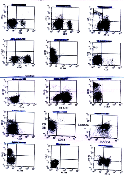 |
Figure 3: Dot plots showing
immunoreactivity pattern of prolymphocytes in B-cell prolymphocytic leukemia |
Prolymphocytic
leukemia was first described in 1974 by Galton, as a rare variant of
chronic lymphocytic leukemia.3-6 It is a rare chronic chronic
lymphoproliferative disorder that includes two subtypes, B cell and
T cell, each with its own distinct clinical, laboratory and pathological
features.4 Most patients are over 60-year old, with a
median age of 65-69 and similar male: female distribution.1
The key features
of B-PLL are massive splenomegaly with absent or minimal peripheral
lymphadenopathy and a rapidly rising lymphocyte count, usually over
100x109/L. Anemia and thrombocytopenia are seen in about half the number
of cases.1,3,7 T-PLL patients usually present with generalized
lymphadenopathy and skin lesions.[1] Morphologically, majority
( >55% and usually >90%) of the circulating cells are prolymphocytes;
that is, medium sized cells (twice the size of a small lymphocyte),
with a round nucleus, moderately condensed nuclear chromatin, a prominent
central nucleolus and a relatively small amount of faintly basophilic
cytoplasm. In contrast, the cells of T-PLL are small to medium-sized
lymphoid cells with agranular basophilic cytoplasm,and have a markedly
irregular nuclei.1,2,8 Typically, B-PLL is differentiated from chronic lymphocytic leukemia [CLL] and CLL/PL with 55% prolymphocytes being a key criteria. B prolymphocytes
are more uniform and have a more regular nuclear outline than those
of CLL/PL. Also, CLL/PL have 10-55% prolymphocytes and a variable number
of plasmacytoid lymphocytes.4 The distinction from Hairy
cell leukemia [HCL] variant is based mainly on the appearances of
the cytoplasm. In HCL variant, the cytoplasm is more abundant and distinctly
villous, whereas in B-PLL it is generally smooth.1,2
The bone marrow
shows interstitial or nodular infiltrate of nucleolated cells with an
intertrabecular distribution.1,8 In the present case,
the bone marrow was diffusely infiltrated by prolymphocytes with marked
suppression of normal hematopoietic elements. The cells of B-PLL strongly
express B-cell antigens such as CD19, CD20, CD22, CD79a and CD79b. In
this case the patient strongly expressed CD19, CD20, CD22, CD23 and
CD45. The cells of T-PLL express CD2,CD3 and CD7.The reported median
survival is 3 to 4 years for patients with prolymphocytic leukemia and
8 years for those with CLL. Patients with T-PLL have even poorer prognosis
than those with B-PLL.1,2,8 B-PLL responds poorly o
therapies for CLL and T-PLL. Hence, typing PLL is very critical for
both therapeutic and prognostic purposes.
In conclusion,
a precise diagnosis by means of a comprehensive approach involving clinical,
morphological and immunophenotypic features are extremely critical in
this era for patient management and prognostication. Also, this case
illustrates the importance of immunophenotyping as an adjunct to morphology
in the diagnosis of chronic lymphoproliferative disorders.
- Campo E,
Catovsky D, Montserrat E, Muller-Hermelink HK, Harris NL, Stein H. B-cell
prolymphocytic leukemia. In: WHO Classification of tumors of hematopoietic
and lymphoid tissues. 4th edn. Lyon 2008. Pg.183-184.
-
Naseem S,
Gupta R, Kashyap R, Nityanand S. T-cell prolymphocytic leukemia: a report
of two cases with review of literature. Indian J. Hematol Blood Transfus
2008;24(4):178-181.
-
Katayama
I, Aiba M, Pechet L, Sullivan J, Robert P, Humphreys RE. B- lineage
prolymphocytic leukemia as a distinct clinicopathologic entity. Am J Pathol 1980;99:399-412.
-
Kar R, Kumar
R, Tyagi S. De-novo CD5+ B-prolymphocytic leukemia presenting at younger
age with favourable outcome. Turk J Hematol 2008;25:149-151.
-
Schlette
E, Bueso-Ramos C, Giles F, Glassman A, Hayes K, Medeiros J. Mature B-cell
Leukemias with more than 55% prolymphocytes ñ A heterogenous group
that includes an unusual variant of Mantle cell lymphoma. Am J Clin
Pathol 2001;115:571-581.
-
Merchant
S, Schlette E, Sanger W, Lai R, Medeiros J. Mature B-cell Leukemias
with more than 55% prolymphocytes ñ Report of 2 cases with Burkitt
Lymphoma ñ type chromosomal translocations involving c-myc. Arch Pathol
Lab Med 2003;127:305-309.
-
Bearman
RM, Pangalis GA, Rappaport H. Prolymphocytic Leukemia ñ Clinical,
Histopathological and Cytochemical Observations. Cancer 1978;42:2360-2372.
-
Nayak KS,
Narayanan S, Naik R, Khadilkar UN. Prolymphocytic Leukemia ñ report
of three cases. Indian J Pathol Microbiol 2003;46(3):459-461.
|

